
Subject
Anatomy
Anatomy is a subject in art. The gallery holds 57 works in this subject in art, including works by Daniel Huntington, Edwin Landseer and Cornelis Troost.
Anatomy (from Ancient Greek ἀνατομή (anatomḗ) 'dissection') is the branch of morphology concerned with the study of the internal and external structure of organisms and their parts. Anatomy is a branch of natural science that deals with the structural organization of living things.
Key artists

Overview
Anatomy, from the Ancient Greek ἀνατομή (anatomḗ) meaning 'dissection,' is the branch of natural science concerned with the internal and external structure of organisms and their parts. As a subject in art, anatomy represents one of the most enduring and consequential intersections of scientific inquiry and visual representation. The study of anatomy became essential to artistic training because the principal subject matter of Renaissance artists was the human body, and to paint it correctly required understanding its underlying structure. This dual nature, scientific and aesthetic, has made anatomical depiction a recurring motif across centuries of art history, from life-size illustrations to delicate paper flaps revealing the body's interior, from woodcut prints to neon installations. The Getty Research Institute's 2022 exhibition 'Flesh and Bones: The Art of Anatomy' explicitly celebrated this connection, noting that artists not only helped create anatomical images but were part of the market for them, as anatomy was a basic component of artistic training for centuries.
Iconography & meaning
The iconography of anatomy in art carries multiple layers of meaning. At its most practical, anatomical imagery served as instruction, maps of the body for medical practitioners and artists alike. The earliest printed anatomical atlases in Europe, introduced during the Renaissance, provided new visual maps to the body, often composed of striking images. Yet anatomical representation also engaged with deeper symbolic registers. The Getty exhibition organized its exploration around six themes that reveal this range: Anatomy for Artists; Anatomy and the Antique; Lifesize; Surface Anatomy; Three Dimensionality; and The Living Dead. The last of these is particularly resonant, examining the motif of the representation of the dead as living, with skeletons and anatomized cadavers capable of motion rather than inert on a dissecting table. Anatomists and artists approached the problem of how best to describe the body's complex and invisible interior with a variety of representational strategies, ranging from the graphic to the sculptural and, recently, the virtual. Paper-flap constructions allowed viewers to lift and peer under layers of flesh, while stereoscopic photographs mimicked binocular perception and projected anatomical structures into space. These strategies cultivated an understanding of anatomy as a synthetic whole rather than merely a collection of parts.
Across movements & periods
The treatment of anatomy in visual art changed dramatically across movements and periods. In antiquity, anatomical knowledge was advanced through dissection, with the first recorded school of anatomy formed in Alexandria from the late fourth century to the second century BCE, where Herophilos and Erasistratus performed human dissection and vivisection. Galen, active in the second century CE, reached the apex of ancient anatomical knowledge through his dissections of animals, and his teachings formed the foundation of medical education throughout the Middle Ages. The Renaissance brought renewed engagement with anatomical dissection, inspired by humanist ideals. Andreas Vesalius (1514-1564) corrected centuries of unquestioned assumptions with his revolutionary publications, becoming the founder of modern anatomy. From the 15th century, anatomical teaching through dissection of human cadavers spread through European universities. The 18th century saw landmarks such as the publications of Albinus, while the 19th and 20th centuries introduced new representational technologies, from photography to digital imaging. The Getty exhibition noted that three-dimensionality was inventively pursued in the pre-digital age, and that recent approaches include the virtual. Leonardo da Vinci's own anatomical studies, begun in the 1480s and pursued in two major campaigns around 1490 and 1506-1513, illustrate this evolution: he dissected perhaps thirty human corpses and many more animals, developing illustrative techniques borrowed from architecture and engineering to convey the three-dimensional form of the body. His intended treatise, had he published it, would have been the single most important work on anatomy ever written.
Notable depictions
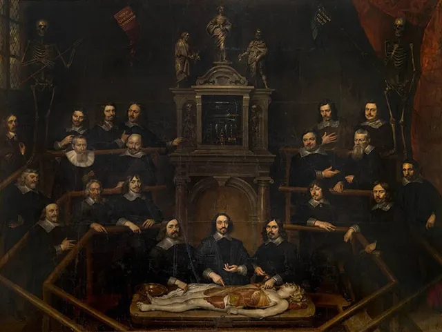
The most celebrated anatomical depictions in art history include Leonardo da Vinci's drawings, which remained unpublished at his death and entered the Royal Collection around 1670 through the collector Pompeo Leoni. Leonardo investigated the nervous system, internal organs, bones and muscles, heart structure and function, and reproductive systems, recording his findings in hundreds of detailed drawings and thousands of words of discussion. His famous 'Vitruvian Man,' showing a man in a square and circle with his body divided into equal portions, represented his search for ideal human proportion. In the Dutch Golden Age, group portraits of anatomy lessons became a significant genre, exemplified by Rembrandt's 'The Anatomy Lesson of Dr. Nicolaes Tulp' and echoed in this collection's 'Anatomy lesson of Dr Willem Röell' and 'The Anatomy Lesson of Dr. Joannes van Buyten.' These works documented the public, civic nature of anatomical knowledge. The 18th-century publications of Albinus represented another landmark, continuing the tradition of spectacular life-size anatomical illustration. More recently, the Getty's 2022 exhibition 'Flesh and Bones' showcased works ranging from a pocket-size book of anatomy for artists from over 200 years ago to contemporary pieces using neon, demonstrating the continuing vitality of anatomical representation across media.
Legacy
The legacy of anatomy in art extends far beyond its historical applications. The Warren Anatomical Museum, founded in 1847 by Harvard professor John Collins Warren, illustrates how anatomical collections became technologies of memory, comparison, and authority. The museum, now part of the Francis A. Countway Library's Center for the History of Medicine, preserves both knowledge and the social relations that made that knowledge possible, including ongoing reckoning with connections to slavery and scientific racism. John Collins Warren's 1831 lobbying for Massachusetts' law legalizing anatomical dissection was the first of its kind in the United States, and his dying request was his own medical dissection. For contemporary practice, gross anatomy has become a key part of visual arts; basic concepts of how muscles and bones function and deform with movement remain essential to drawing, painting, or animating the human figure. The Getty Research Institute's emphasis on the 'shared vocabulary of anatomical images' and the 'different methods used to reveal the body through a wide range of media, from woodcut to neon' underscores this continuity. Leonardo da Vinci's anatomical drawings, finally published and translated in the late 19th century, are now understood as the work of one of the greatest anatomists ever to have lived, whose illustrative challenges were in many ways the same as those faced by anatomists today.
Works
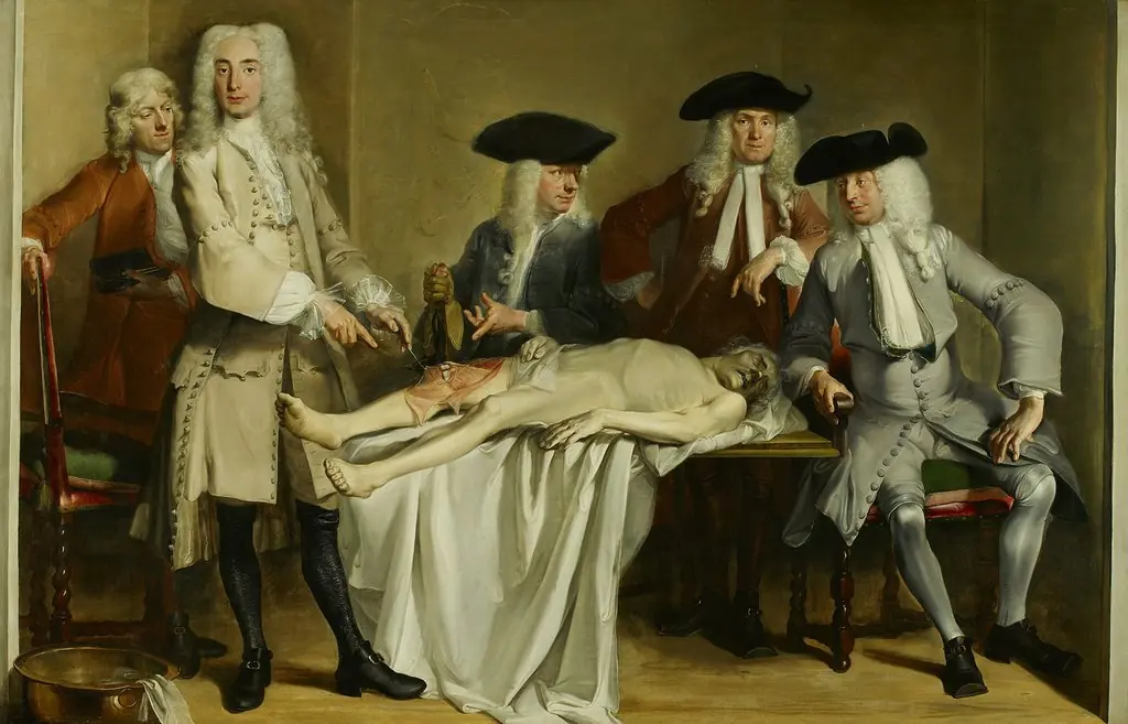 Anatomy lesson of Dr Willem Röell.
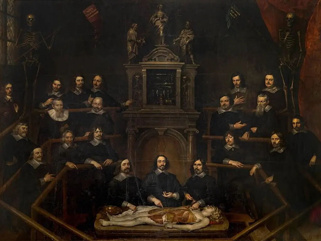 The Anatomy Lesson of Dr. Joannes van Buyten
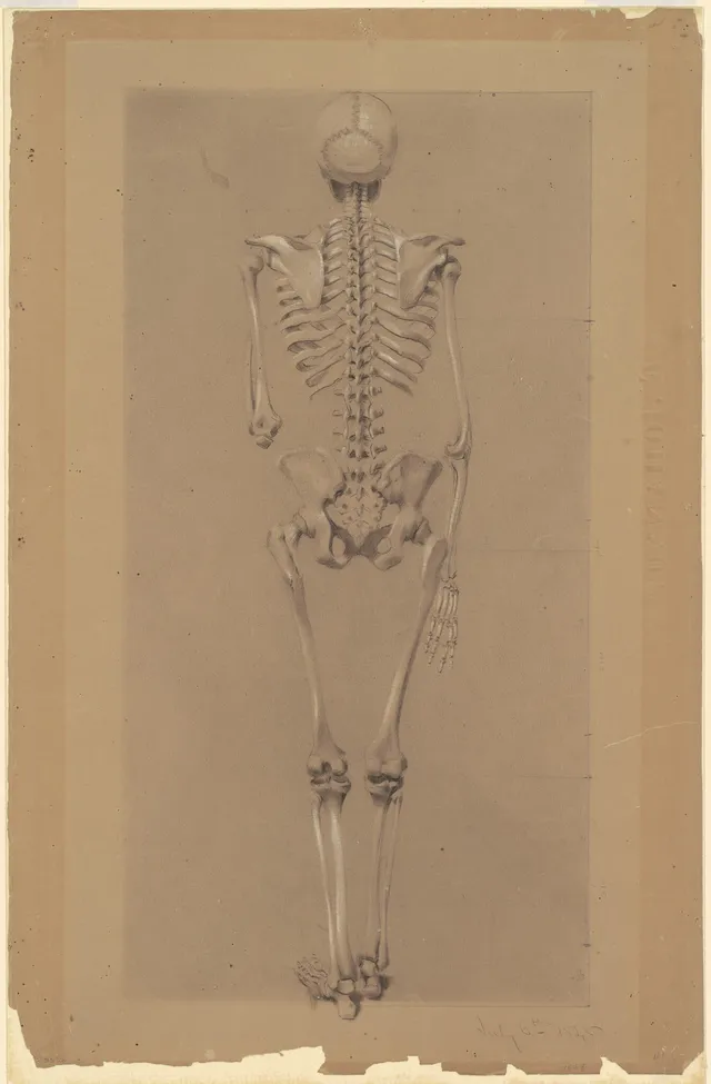
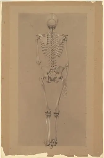 Rear View of a Skeleton
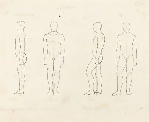 Extent of Motion, Front and Side View Equipoised, Supported on One Leg
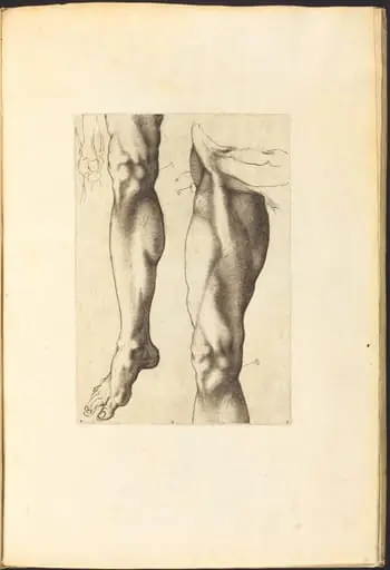 Print from Drawing Book
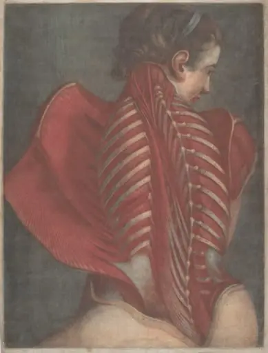 L'Ange anatomique (The Anatomical Angel) or Dissection of a Woman's Back
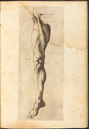 Print from Drawing Book
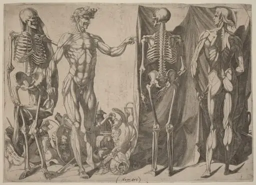 Squelettes et Ecorches
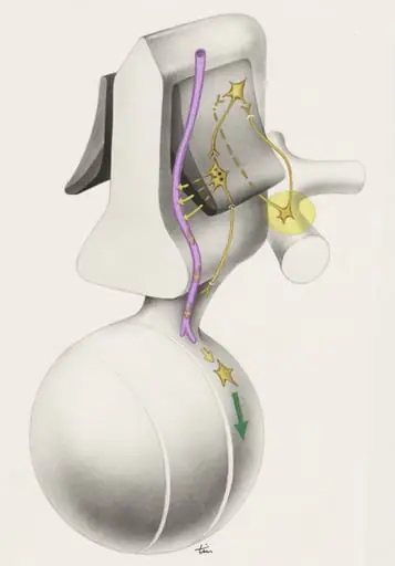 Part of the central nervous system
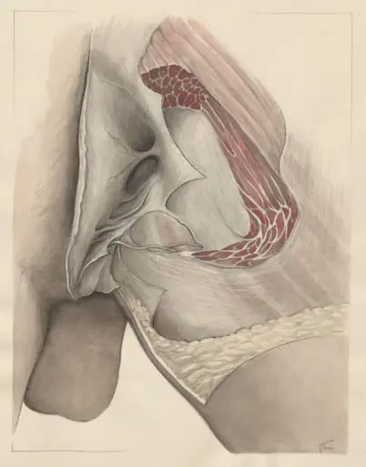 Tissue of the buttocks
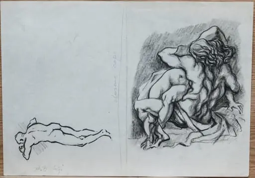 Desene copii
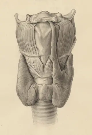 Glands of internal secretion
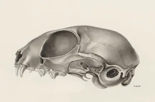 Skull of a feline
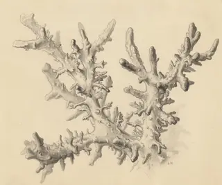 Part of the human anatomy
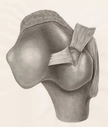 Muscular tissue on a joint
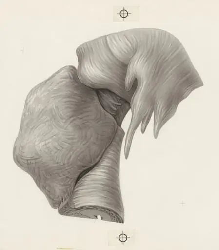 Human tissue
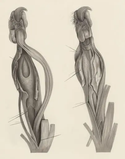 Muscular tissue of an animal paw
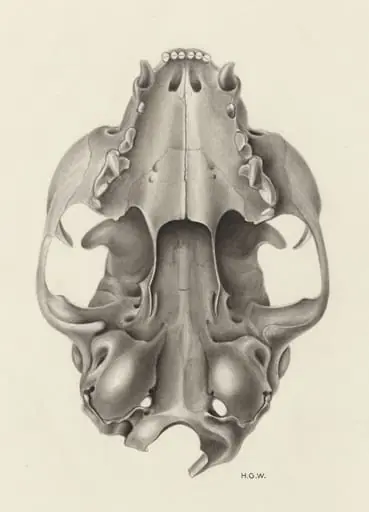 Skull of a feline seen from below
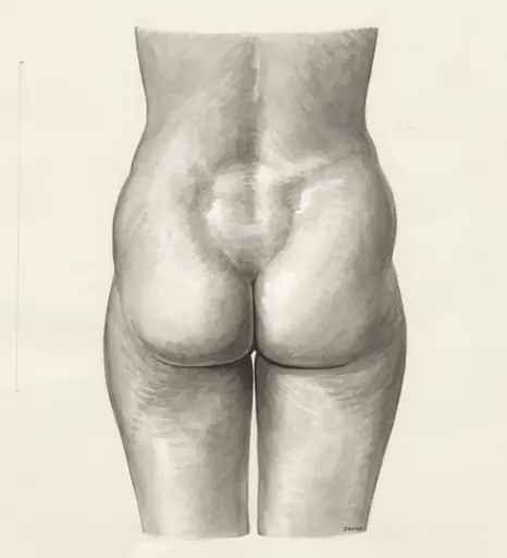 Female lower back and buttocks
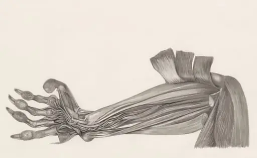 Muscular tissue of an animal arm
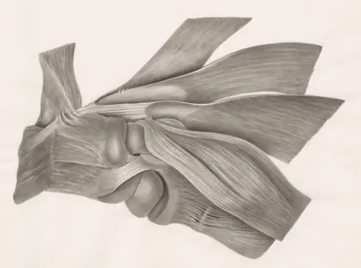 Muscular tissue
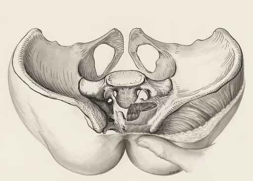 Cross section of the pelvis
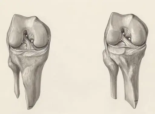 Muscular tissue of a joint
 Female torso, holding up the right arm
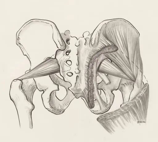 Muscular tissue of the pelvis
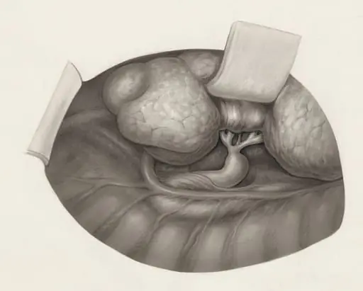 Human organs
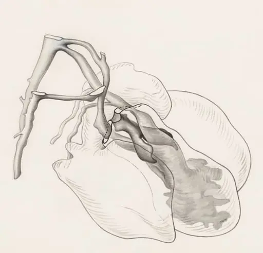 Tissue of the human embryo
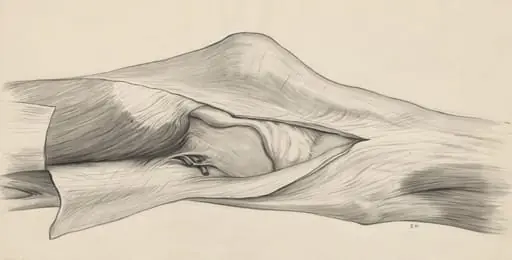 Knee joint
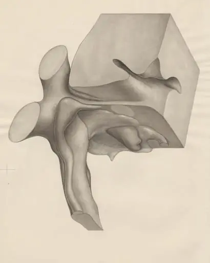 Undefined tissue
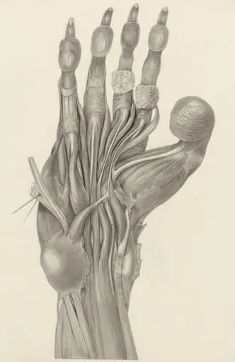 Muscular tissue of the claw of a mammal
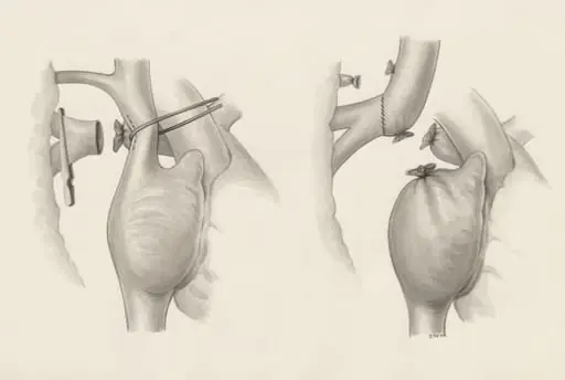 Heart surgery
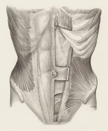 Muscular tissue of the abdomen
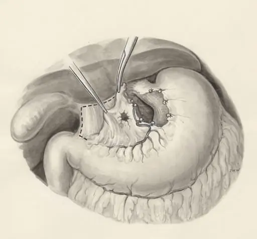 Surgery on a part of the human body
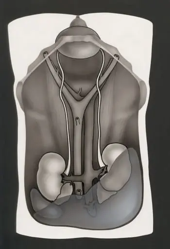 Opened abdominal cavity, with the kidneys
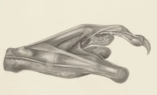 Muscular tissue of the claw of a gray four-eyed opossum
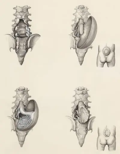 Protrusion of the lower back
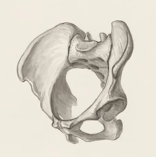 Pelvis bones
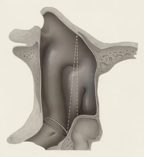 Respiratory system
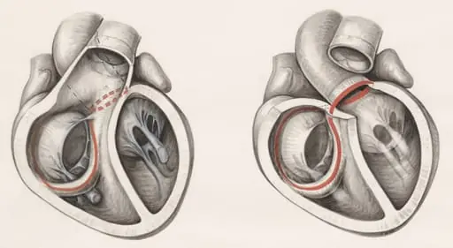 Two cross sections of the human heart
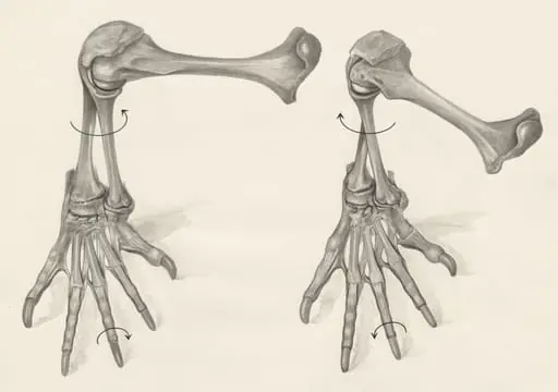 Skeletal legs of a lizard
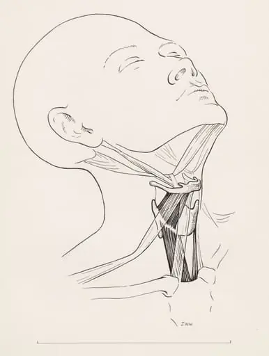 Muscular tissue of the human neck
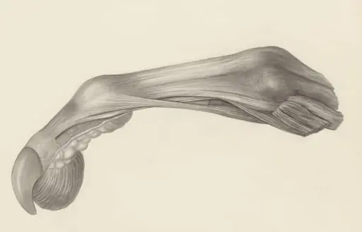 Muscular tissue of the finger of a rhesus macaque
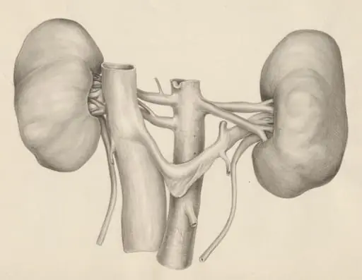 Kidneys
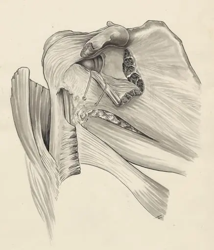 Damage of the pelvic bones
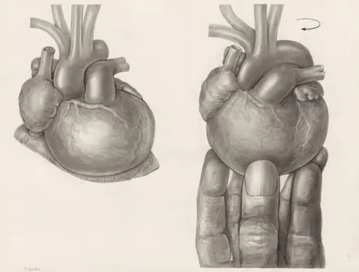 Two hearts, one being rotated by a hand
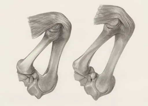 Muscular and bone tissue of the hand or fingers
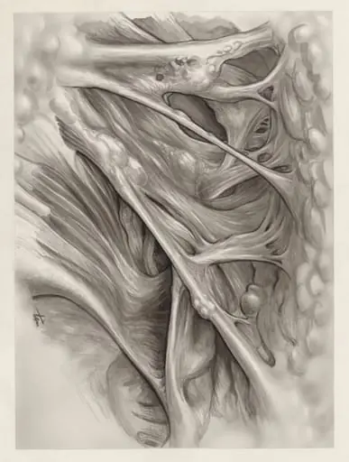 Muscular tissue of the hand or fingers
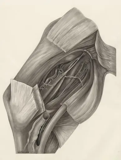 Muscular tissue of the hand or fingers
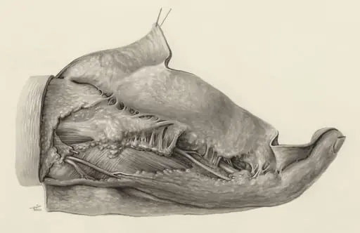 Muscular tissue of the hand and finger
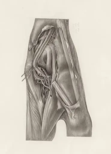 Muscular tissue of the upper extremities
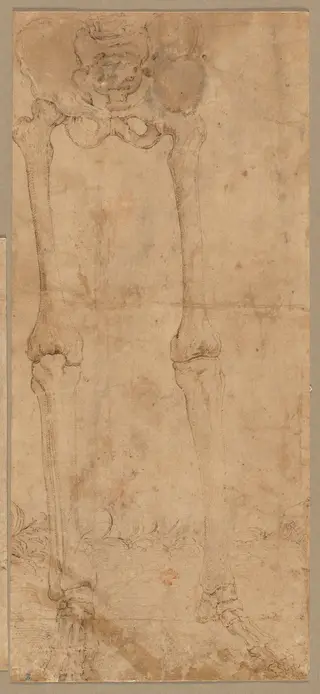 Lower Half of Skeleton from the Front
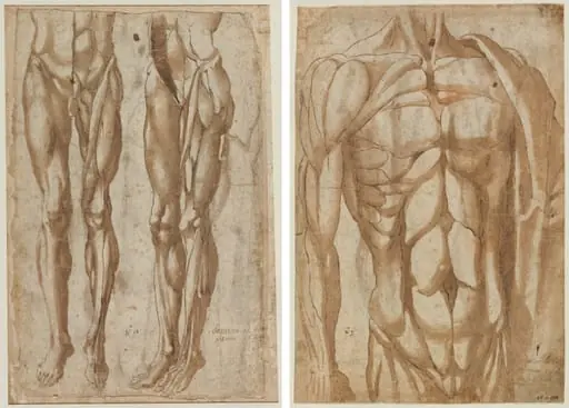 Two Studies of a Flayed Man (recto) Study of a Flayed Torso (verso)
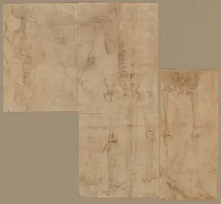 Skeleton
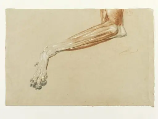 Ecorche drawing of a dog's leg
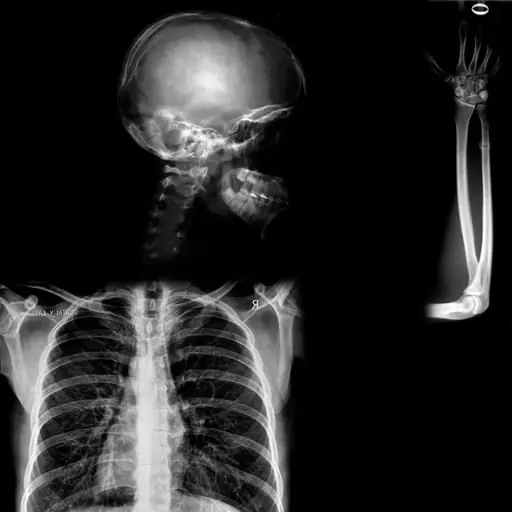 Άτιτλο
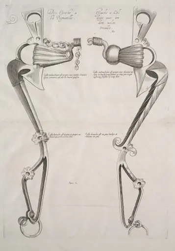 Diagram for Horse's Bit
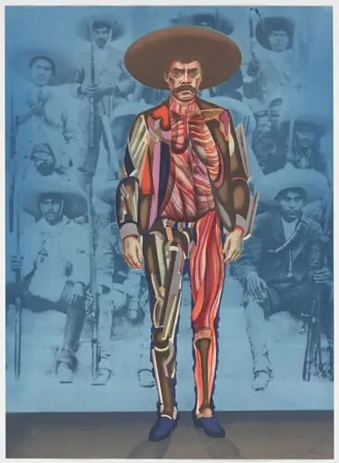 Untitled
Frequently asked questions
What is Anatomy?
Anatomy (from Ancient Greek ἀνατομή (anatomḗ) 'dissection') is the branch of morphology concerned with the study of the internal and external structure of organisms and their parts.
How many Anatomy works does Artifact World Gallery have?
Artifact World Gallery holds 57 public-domain Anatomy works, all free to view and download.
Who are the key Anatomy artists?
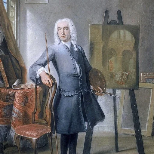
Key Anatomy artists in the collection include Daniel Huntington, Edwin Landseer and Cornelis Troost.
Where can I see Anatomy works?
Anatomy works in the collection are held by Leiden University Libraries, National Gallery of Art and Cleveland Museum of Art.